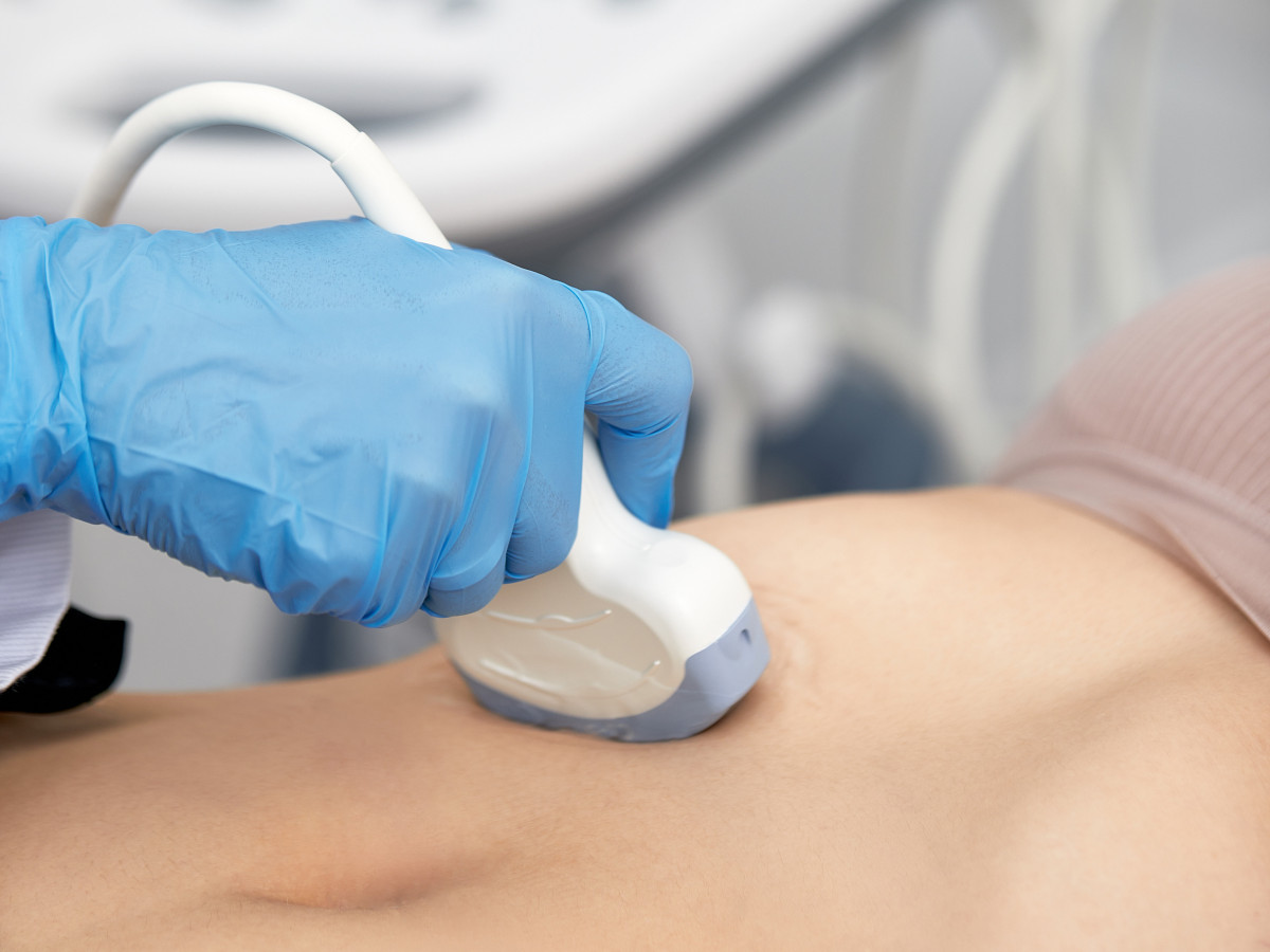
Elastography – Benefits and function
Some diseases such as liver cirrhosis or certain tumors lead to changes in the tissue. Tissue differentiation is necessary in order to be able to make specific diagnoses. Conventional imaging methods can only fulfill to a limited extent, which is why (ultrasound) elastography has increasingly turned out to be the favored method. Sonoelastography is able to show tissue stiffness non-invasively and repeatably. There are many different techniques available to the treating physicians.
In the following article we will tell you what constitutes elastography and what benefits it offers for medical technology design.
What does elastography mean?
Elastography is a non-invasive and risk-free imaging method that can precisely measure and display the stiffness or elasticity and viscosity of tissue. Just like conventional scanning examinations, elastography takes advantage of the fact that tumor tissue is firmer and more dense than healthy tissue. In contrast to manual palpation, however, the examination results here do not depend on the doctor's subjective sense of touch, but are objective and reliable. The doctor can easliy diagnose the solid tumor tissue by highlighting it with color in the elastogram. Stiff tissue is shown on the elastogram in blue or violet, medium-hard tissue in yellow or green and soft tissue in red. Elastography shows the stiffness of the soft tissue by measuring the degree of deformation under pressure.

Elastography areas of application
A change in tissue elasticity is caused by various diseases. In addition to liver diseases, these above all include tumors or calcification in connection with arteriosclerosis. The clinical benefit of elastography is therefore, on the one hand, in the early detection and differential diagnosis of diseases, since it can reflect the changes. Furthermore, it improves the diagnosis of fibrosis diseases such as cancer or chronic hepatitis through the extent of the lesions and their expansion. In addition, elastography makes it easier to assess response to treatments such as chemotherapy or radiofrequency ablation.
In addition, ultrasound elastography can also be helpful in finding the cause of chronic pain in the musculoskeletal system. This way, even the hardening of the muscles and fascia can be made visible, in contrast to conventional imaging methods.
Strain elastography and shear wave elastography
Elastography displays the stiffness of soft tissue. Depending on the structure of the mechanical compression, a distinction is made between two different techniques: Strain elastography and shear wave elastography.
- With strain elastography, the mechanical force required to deform the tissue is applied by the user themselves. The external pressure is generated by actuating the transducer at the desired body site using cardiovascular pulsation or breathing movements. The less the stretch, the harder the lesion and vice versa. Strain elastography is therefore also referred to as strain imaging. A disadvantage of this method is that the applied compression pressure cannot be measured exactly. Thus, the absolute tissue strain cannot be adequately calculated.
- Shear wave elastography, on the other hand, uses a shear wave to trigger mechanical stress. The propagation speed is then measured by ultrasound. This shear wave, in turn, can be caused by 2 different methods in the form of acoustic radiation force impulses or by external mechanical vibration.
Magnetic resonance elastography
Magnetic resonance elastography can be used to display an elastogram that reflects the stiffness of body tissue. Unlike the methods described above, magnetic resonance elastography is not performed with an ultrasound device, but with an MRI device. The main purpose of this type of elastography is to diagnose stiffening of the liver in chronic liver disease.
Traditionally, liver biopsy has often been used for this. However, magnetic resonance elastography offers a number of advantages over this:
- non-invasive and safer
- the entire liver can be assessed and not just parts of the tissue
- a fibrosis can be detected earlier
- magnetic resonance elastography is particularly suitable for overweight people
- the risk of certain liver complications can be predicted
Magnetic resonance elastography supplements the cheaper and easier-to-perform fibroscan in the diagnosis of liver fibrosis. However, one cannot replace the other. Rather, the elastography is consulted as an additional method to examine the previously determined liver stiffness more precisely. Magnetic resonance elastography is not suitable for people who wear metallic or electronic devices, as these can produce image errors. In addition to jewellery, these include pacemakers, metal joint prostheses, etc.

Elastoghraphy – improving the usability of products
The handling of equipment for elastography is particularly important for treating physicians. UX medical technology describes a part of our work that deals with improving the user experience when operating instruments. In the best case, devices are prepared in such a way that they can be used intuitively. This noticeably increases usability and minimizes treatment errors.
Do you also want to bring a new medical product onto the market? Then benefit from a formative evaluation in which your device is put through its paces by real users.
If you have any further questions about elastography or other topics related to medical technology design, please feel free to contact us at any time. We look forward to your inquiry and will reply to you as soon as possible.
Read next:
