Analog X-ray - Medical imaging processes
Analog X-ray is an imaging method in medicine that provides diagnostic information about bones, internal organs or blood vessels. However, the procedure is considered outdated in medical design and has now been almost completely replaced by the innovative equivalent of digital X-rays. Due to its revolutionary power, it is still worth exploring the methodology behind today's analog X-rays. Furthermore, the question arises, what the advantages of digital X-rays are and how old X-ray images and devices can be disposed of.
In our new blog post, we have summarized everything you need to know about analog X-rays.
What is analog X-ray?
The term "analog X-ray" refers to the image receiver system of an X-ray facility. This analogue image detector consists of three components: the X-ray film, the intensifying screen and the lighproof housing. The storage film used for analogue X-rays consists of a fine, flexible and transparent polyester film and a thin layer of photographic emulsion. This emulsion, in turn, is coated with protective polymer layers and a matting agent. However, in addition to the X-ray film developer and the X-ray chemicals, a water bath is required for the development of the analogue X-ray image.

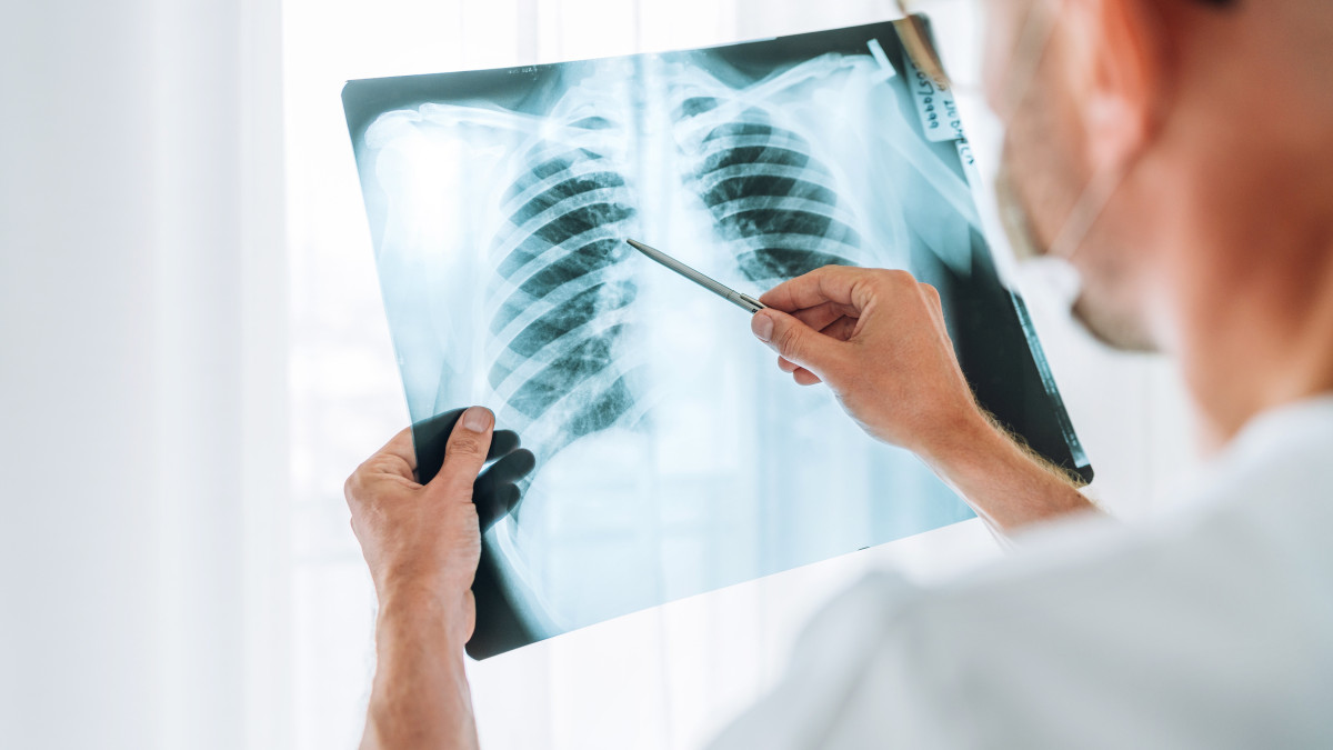
Analog X-ray: The process
After the X-rays are emitted by the X-ray machine and partially absorbed by the patient's body, they hit the X-ray film. Now the process of analog X-ray image development begins. As soon as the X-ray film is exposed to X-rays, so-called black silver develops from the silver bromide crystals of the storage film and an image is created. In the developer bath, further black silver is formed from the silver bromide crystals that are still partially present. In analog X-rays, the remaining silver bromide that has not yet been exposed is then dissolved out in the fixing bath and black silver remains in the areas that were not exposed by the X-ray film. With an imaging plate scanner, the analog X-ray imaging plates can then be digitized, which is particularly advantageous for storing the images.
Analog X-ray: Regular consinstency check-ups
X-ray systems with analog X-ray detectors are subject to consistency tests that must be carried out regularly. The basis for this are §§ 16 X-ray Ordinance (RöV), guidelines for the implementation of quality assurance in X-ray facilities for the examination or treatment of people according to §§ 16 and 17 X-ray Ordinance, quality assurance guidelines (QS-RL) and DIN standards (DIN 6868-2, DIN 6868-5). The radiation protection officers are responsible for the regular constancy tests. Constancy tests must be documented and, if necessary, made accessible to the respective X-ray departments.
Analog X-ray: Outdated technology with modern alternatives
As already mentioned at the beginning, digital X-rays have more advantages than disadvantages compared to analog X-rays. This is one of the reasons why the vast majority of people today rely on their digital counterpart. Analog X-rays are not only more expensive, they also take significantly longer. In addition, a higher dose of radiation that is hazardous to the patient's health is emitted, which is no longer required today. The results from analog X-rays also have poorer image quality.
In contrast to analog X-rays, digital X-rays use special imaging plates or sensors instead of exposed film material . The high sensitivity of the imaging plate during radiography makes it possible to obtain high-resolution images with up to 90% less radiation. Instead of conventional analog X-ray images, which are later created with chemicals in a darkroom, digital X-ray images are received by a special reader and stored directly on a computer. This means that digital X-rays offer a clear advantage over analog X-rays, especially when it comes to usability. A further development of this and thus in stark contrast to analog X-rays is digital direct radiography. In this case, the X-rays are guided directly into a detector without first passing through an imaging plate system. This detector converts the X-rays directly into electronic image information and the findings are available on the PC within a few seconds.
Disposal of analog X-ray images
Analog X-rays are not only being phased out because of technical implementation issues, but also for reasons of environmental protection. This is because analog X-ray images are significantly more harmful to the environment than image plates, which can be overwritten and reused up to 100 times. Nevertheless, the phosphor contained in it is partly made of toxic barium fluoride. Barium is also a heavy metal. Therefore, these fluorescent panels must be disposed of as hazardous waste according to waste code AVV 09 01 99.
Theoretically, private individuals can dispose of analog X-ray images in the household or residual waste, but this is not recommended for reasons of environmental protection and recycling. Recycling old X-rays can recover more than 90% of raw materials, including precious silver. About 2.5 kg of silver can be obtained from one ton of X-ray film material, as well as various plastics and paper components. If you dispose of analog X-ray images in the household waste, then no raw materials can be recycled. It is therefore recommended that the analog X-ray images be handed in at the doctor's office or hospital where they were created. They are required by law to take them back. It is also possible to drop them off at a recycling center or disposal company nearby. There are now also a number of specialized companies that collect the analog X-rays from your home.
If you have any further questions about analog X-rays, please feel free to contact us at any time. We look forward to your inquiry and will reply as soon as possible.
Read next: